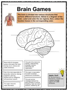
40 diagram of the brain with labels and functions
Anatomy and Function of the Human Brain - Study.com A human brain is composed of several parts, each with its own function. The parts of the brain include the cerebrum, the cerebellum, the brain stem, and the pituitary gland. The brain structure is ... Achiever Papers - We help students improve their academic standing All our academic papers are written from scratch. All our clients are privileged to have all their academic papers written from scratch. These papers are also written according to your lecturer’s instructions and thus minimizing any chances of plagiarism.
› cold-and-flu › ear-infectionPicture of the Ear: Ear Conditions and Treatments - WebMD The spiral-shaped cochlea is part of the inner ear; it transforms sound into nerve impulses that travel to the brain. The fluid-filled semicircular canals (labyrinth) attach to the cochlea and ...

Diagram of the brain with labels and functions
Brain: Anatomy, Pictures, Functions, and Conditions - Verywell Mind The midbrain controls many important functions such as the visual and auditory systems as well as eye movement. 4. Portions of the midbrain called the red nucleus and the substantia nigra are involved in the control of body movement. The darkly pigmented substantia nigra contains a large number of dopamine-producing neurons. Help:Displaying a formula - Wikipedia Care should be taken when writing sets within {{}}, as braces, equal signs, and vertical bars can conflict with template syntax.The {{}} template is available for braces, as shown in the example above.Likewise, {{}} encloses its parameter inside vertical bars to help with the pipe character conflicting with template syntax.For a single vertical bar, use {{}}, and for an equal sign, use {{}}. nervous system part 2 HW mylab Flashcards | Quizlet Drag the labels from the left onto the appropriate targets in the diagram on the right to describe the functions of the labeled brain structures. Not all labels will be used. a. serves as sophisticated center of integration and control b. controls the left side of the body
Diagram of the brain with labels and functions. Eye Diagram With Labels and detailed description - BYJUS The optic nerve transmits electrical signals from the retina to the brain. Pupil is the opening at the centre of the iris. Its size changes with the amount of light. The retina lines the back of the eye and contains several photoreceptors. Vitreous humour is the fluid present in the centre of the eye and provides form and shape to the eye. achieverpapers.comAchiever Papers - We help students improve their academic ... All our academic papers are written from scratch. All our clients are privileged to have all their academic papers written from scratch. These papers are also written according to your lecturer’s instructions and thus minimizing any chances of plagiarism. byjus.com › biology › diagram-of-eyeEye Diagram With Labels and detailed description - BYJUS The optic nerve transmits electrical signals from the retina to the brain. Pupil is the opening at the centre of the iris. Its size changes with the amount of light. The retina lines the back of the eye and contains several photoreceptors. Vitreous humour is the fluid present in the centre of the eye and provides form and shape to the eye. Brain: Function and Anatomy, Conditions, and Health Tips The brain is an organ that's made up of a large mass of nerve tissue that's protected within the skull. It plays a role in just about every major body system. Some of its main functions ...
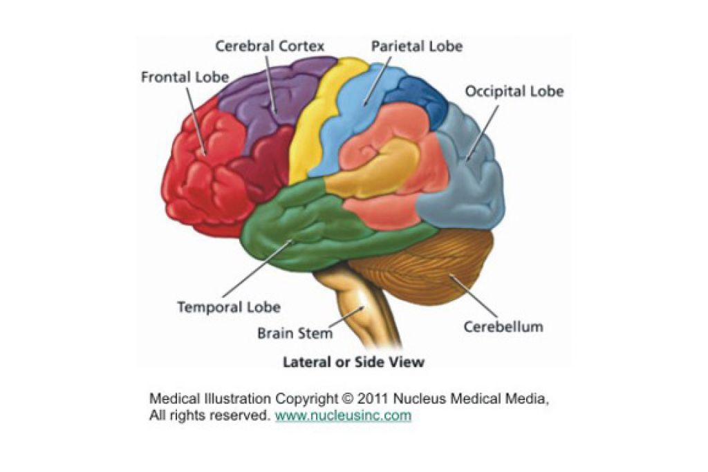
Ventricles of the Brain: Anatomy, Function, CSF Flow - EZmed Learn the ventricles of the brain along with their definition, function, location, anatomy, and cerebrospinal fluid (CSF) flow using labeled diagrams. The ventricular system contains the lateral, third, and fourth ventricles whose function is to produce cerebrospinal fluid. Learn where CSF is found, Labeled Diagrams of the Human Brain You'll Want to Copy Now Labeled Diagrams of the Human Brain Central Core The central core consists of the thalamus, pons, cerebellum, reticular formation and medulla. These five regions are the central areas that regulate breathing, pulse, arousal, balance, sleep and early stages of processing sensory information. Brain (Human Anatomy): Picture, Function, Parts, Conditions ... - WebMD • The cerebellum is at the base and the back of the brain. The cerebellum is responsible for coordination and balance. The brain is also divided into several lobes: • The frontal lobes are... Diagram Of Brain with their Labelings and Detailed Explanation - BYJUS A well-labelled diagram of a human brain is given below for further reference. Structure And Function Of The Human Brain Parts Of The Human Brain The human brain is divided into three main parts: Forebrain. Midbrain. Hindbrain. These three main parts comprises many small parts. Forebrain The forebrain is also called as Prosencephalon.
28+ Human Brain Diagram With Labels And Functions Pictures Brain diagram labeled with functions. It is made up of more than 100 billion nerves that communicate in trillions of. Learn about parts of the brain and interesting facts about the the brain weighs just about two to three pounds and appears like a walnut. The frontal lobe, parietal lobe, temporal lobe, occipital lobe, cerebellum, and brainstem. byjus.com › biology › diagram-of-heartHeart Diagram with Labels and Detailed Explanation - BYJUS The diagram of heart is beneficial for Class 10 and 12 and is frequently asked in the examinations. A detailed explanation of the heart along with a well-labelled diagram is given for reference. Well-Labelled Diagram of Heart. The heart is made up of four chambers: The upper two chambers of the heart are called auricles. Picture of the Ear: Ear Conditions and Treatments - WebMD WebMD's Ear Anatomy Page provides a detailed image and definition of the ear as well as an overview of ear-related health problems. Learn about the ear's function in the body and test and ... A Labelled Diagram Of Neuron with Detailed Explanations - BYJUS A Labelled Diagram Of Neuron with Detailed Explanations Biology Biology Article Diagram Of Neuron Diagram Of Neuron A neuron is a specialized cell, primarily involved in transmitting information through electrical and chemical signals. They are found in the brain, spinal cord and the peripheral nerves. A neuron is also known as the nerve cell.
Brain Jack Image: Brain Function Chart
Human Brain - Structure, Diagram, Parts Of Human Brain - BYJUS The hypothalamus is a small and essential part of the brain, located precisely below the thalamus. It is considered the primary region of the brain, as it is involved in the following functions: Receives impulses Regulates body temperature Controls the mood and emotions Controls the sense of taste and smell Synthesises the body's essential hormones
Brain Anatomy And Function - Anatomy Drawing Diagram
brain stem diagram labeled Label Parts Of The Brain - Pensandpieces pensandpieces.blogspot.com. brain parts label human regions illustration body. In The Anatomy Of Humans And Of Many Other Vertebrates, The Brainstem . brain anatomy parts human functions diagram function many labels humans stem structure brainstem posterior physiology
Brain Diagram - Cliparts.co
Lobes of the brain: Structure and function | Kenhub The lobes of the cerebrum are actually divisions of the cerebral cortex based on the locations of the major gyri and sulci. The cerebral cortex is divided into six lobes: the frontal, temporal, parietal, occipital , insular and limbic lobes. Each lobe of the cerebrum exhibits characteristic surface features that each have their own functions.
Tim van de Vall - Comics & Printables for Kids
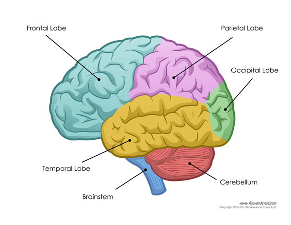
Lobes of the Brain: Cerebral Cortex Anatomy & Function - EZmed The cerebral cortex has 4 main lobes - frontal lobe, parietal lobe, occipital lobe, and temporal lobe - and their location, function, and anatomy all differ. We will use labeled diagrams and lateral images of the brain (side views) to walk through each lobe of the cerebrum. Every EZmed post is filled with simple tricks to remember the content ...

Educative diagrams: The middle of the Brain
The Human Brain (Diagram) (Worksheet) | Therapist Aid The Human Brain (Diagram) The Human Brain Diagram is a versatile tool for psychoeducation. The diagram separates the brain into six major parts, and provides a brief description of the functions carried out by each section. Discussion of the brain, and how it works, can be a powerful way to explore many topics.
The Human Body Facts, Worksheets & Key Systems For Kids
coursehelponline.comCourse Help Online - Have your academic paper written by a ... All our academic papers are written from scratch. All our clients are privileged to have all their academic papers written from scratch. These papers are also written according to your lecturer’s instructions and thus minimizing any chances of plagiarism.

33 Label The Bone Model - Labels Database 2020
Labeled Brain Model Diagram | Science Trends The cerebrum is the largest and most complex portion of the human brain. The cerebrum's function is to control our actions and thoughts, either conscious or unconscious, and responses to stimuli. The cerebrum itself is typically divided into four different lobes: the temporal lobe, the parietal lobe, the occipital lobe, and the frontal lobe.

Neuroanatomy - Important brain structures | Study help | Pinterest | Brain structure, Brain and ...
DOC BRAIN ANATOMY FUNCTION CHEAT SHEET - Winston-Salem/Forsyth County Schools Memory (remembering and learning) Amygdala Emotion (aggression) rage, fear Kluecer& Bucy Lesion monkey brain Hypothalamus Regulates thirst, hunger, body temperature, sexual behavior (hormone release). Controls/regulates maintenance reflexes (eating), Homeostasis linked to emotion. Helps govern endocrines. Monitors glands. Controls hunger.

Neurological bases of Communication - Anatomy
Welcome to Butler County Recorders Office Copy and paste this code into your website. Your Link Name

7 best Brain images on Pinterest | Brain anatomy, The brain and Central nervous system
Anatomy of the Brain: Structures and Their Function - ThoughtCo The forebrain is the division of the brain that is responsible for a variety of functions including receiving and processing sensory information, thinking, perceiving, producing and understanding language, and controlling motor function. There are two major divisions of forebrain: the diencephalon and the telencephalon.

Detailed Labeled Diagram Of The Brain - Aflam-Neeeak
PDF Psychology Brain Structure/Anatomy and Function - Talent Middle School Psychology - Brain Structure/Anatomy and Function BRAIN FACTS Composition of the brain: 78% water, 12% lipids, 8% protein, 1% carbs, 2% soluble organics, and 1% salt ... Some products are also labeled incorrectly." Nicotine in e-cigarettes raise blood pressure. Compared to nonusers, users of e-cigarettes have a 71% higher risk of stroke, 59 ...

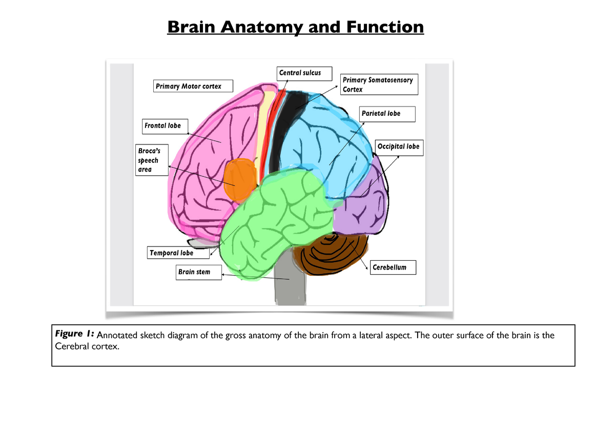
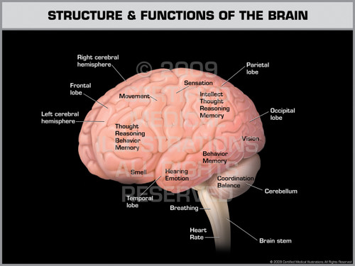
In this diagram it shows the different parts of the brain, as well as the functions they perform ...
Course Help Online - Have your academic paper written by a … 100% money-back guarantee. With our money back guarantee, our customers have the right to request and get a refund at any stage of their order in case something goes wrong.

Diagram of the Brain Quiz
en.wikipedia.org › wiki › Help:Displaying_a_formulaHelp:Displaying a formula - Wikipedia 1) Later on, the text can refer to this equation by its number using syntax like this: As seen in equation ({{EquationNote|1}}), example text... The result looks like this: As seen in equation (1), example text... The equation number produced by {{ EquationNote }} is a link that the user can click to go immediately to the cited equation. Alphabets and typefaces See also: Wikipedia:LaTeX ...
Structure & Functions of the Brain
Botulinum toxin - Wikipedia Botulinum toxin (BoNT), often shortened to Botox, is a neurotoxic protein produced by the bacterium Clostridium botulinum and related species. It prevents the release of the neurotransmitter acetylcholine from axon endings at the neuromuscular junction, thus causing flaccid paralysis. The toxin causes the disease botulism.The toxin is also used commercially for …

Diagram of the Brain and its Functions - Bodytomy Given below is a diagram outlining the main brain functions and parts. Vertical Section of the Brain and its Functions Midbrain The midbrain is divided into two parts by the Aqueduct of Sylvius, which is the duct that connects the IIIrd ventricle in the midbrain with the IV ventricle in the pons and medulla oblongata.

The Human Brain (Diagram) (Worksheet) | Therapist Aid | Human brain diagram, Brain diagram ...
Brain Anatomy and How the Brain Works | Johns Hopkins Medicine The cerebellum ("little brain") is a fist-sized portion of the brain located at the back of the head, below the temporal and occipital lobes and above the brainstem. Like the cerebral cortex, it has two hemispheres. The outer portion contains neurons, and the inner area communicates with the cerebral cortex.

282 best images about Diagramatically Speaking on Pinterest | Respiratory system, Neurons and ...
byjus.com › biology › skin-diagramSkin Diagram with Detailed Illustrations and Clear Labels - BYJUS Skin Diagram The largest organ in the human body is the skin, covering a total area of about 1.8 square meters. The skin is tasked with protecting our body from external elements as well as microbes.

Brain anatomy, Medicine and The o'jays on Pinterest
15.2 Types of Presentation Aids – Stand up, Speak out Charts are also useful when you are trying to explain a process that involves several steps. The two visual aids in Figure 15.8 “Steps in Cell Reproduction” both depict the process of cell division called mitosis using a sequence-of-steps chart, but they each deliver different information. The first chart lacks labels to indicate the different phases of cell division.
Post a Comment for "40 diagram of the brain with labels and functions"