39 brain mri with labels
Arterial spin labeling MRI: clinical applications in the brain Visualization of cerebral blood flow (CBF) has become an important part of neuroimaging for a wide range of diseases. Arterial spin labeling (ASL) perfusion magnetic resonance imaging (MRI) sequences are increasingly being used to provide MR-based CBF quantification without the need for contrast administration, and can be obtained in conjunction with a structural MRI study. brain anatomy | MRI coronal brain anatomy | free MRI cross sectional ... ELBOW AXIAL. WRIST AXIAL. WRIST CORONAL. KNEE CORONAL. KNEE SAGITTAL. ARTERIES UPPER LEG. ARTERIES LOWER LEG. This MRI brain coronal cross sectional anatomy tool is absolutely free to use. Use the mouse scroll wheel to move the images up and down alternatively use the tiny arrows (>>) on both side of the image to move the images.
Enhancing the REMBRANDT MRI collection with expert segmentation labels ... Pyradiomics 41, an open-source python package was used to extract radiomics features from the segmented labels of the MRI brain scans. It included a total of 120 features, ...
Brain mri with labels
101 Labeled Brain Images and a Consistent Human Cortical Labeling ... Another important application of labeled brain images is to train, test, and evaluate automated registration, segmentation, parcellation, and labeling algorithms. ... Automatic anatomical brain {MRI} segmentation combining label propagation and decision fusion. Neuroimage 33, 115-126 10.1016/j.neuroimage.2006.05.061 ... CaseStacks.com - MRI Brain Anatomy Labeled scrollable brain MRI covering anatomy with a level of detail appropriate for medical students. Show/Hide Labels. MRI Brain Anatomy. Back to Anatomy Overview. ... Labelled radiographs and CT/MRI series teaching anatomy with a level of detail appropriate for medical students and junior residents. Pelvis. Pelvic MRI anatomy MRI Brain Atlas - University of Minnesota This web app Atlas is intended for veterinary students and radiologists seeking quick access to canine brain anatomy through a mobile device. Via a toggle button, either MRI images or approximately comparable Brain Transection images may be viewed with or without labels. Navigation & Labels.
Brain mri with labels. Brain MRI: What It Is, Purpose, Procedure & Results - Cleveland Clinic A brain MRI (magnetic resonance imaging) scan, also called a head MRI, is a painless procedure that produces very clear images of the structures inside of your head — mainly, your brain. MRI uses a large magnet, radio waves and a computer to produce these detailed images. It doesn't use radiation. › en › e-AnatomyCross-sectional anatomy of the brain - e-Anatomy - IMAIOS Apr 15, 2022 · Axial MRI Atlas of the Brain. Free online atlas with a comprehensive series of T1, contrast-enhanced T1, T2, T2*, FLAIR, Diffusion -weighted axial images from a normal humain brain. Scroll through the images with detailed labeling using our interactive interface. Perfect for clinicians, radiologists and residents reading brain MRI studies. › 2013 › 07CPT Code for MRI Brain, Breast, Lumbar Spine and Shoulder Find below the latest Radiology CPT codes for for MRI of Brain, Breast, Lumbar Spine and Shoulder: CPT Codes for MRI Lumbar spine In human Lumbar spine is represented by the 5 vertebrae in between the ribcage and the pelvis forming the largest segment of the vertebral column. Depending on the condition that one is treated on these parts of the ... MRI anatomy | free MRI axial brain anatomy - Mrimaster.com This MRI brain cross sectional anatomy tool is absolutely free to use. Use the mouse scroll wheel to move the images up and down alternatively use the tiny arrows (>>) on both side of the image to move the images.
MRI brain (summary) | Radiology Reference Article - Radiopaedia MRI brain is a specialist investigation that is used for the assessment of a number of neurological conditions. It is the main method to investigate conditions such as multiple sclerosis and headaches, and used to characterize strokes and space-occupying lesions. Reference article › en › e-AnatomyNormal chest MDCT with anatomic labels | e-Anatomy - IMAIOS Mar 10, 2022 · IMAIOS and selected third parties, use cookies or similar technologies, in particular for audience measurement. Cookies allow us to analyze and store information such as the characteristics of your device as well as certain personal data (e.g., IP addresses, navigation, usage or geolocation data, unique identifiers). Labeled imaging anatomy cases | Radiology Reference Article ... This article lists a series of labeled imaging anatomy cases by body region and modality. Brain CT head: non-contrast axial CT head: non-contrast coronal CT head: non-contrast sagittal CT head: angiogram axial CT head: angiogram coronal CT... UCLA Brain Mapping Center - ICBM Template To view both the structural MRI and the labels launch the program typing Display icbm_template.mnc -label icbm_labels_corrected.mnc. The opacity of the labels can be set in the Colour Coding menu. The number of each label appears at the bottom left of the orthogonal views window.
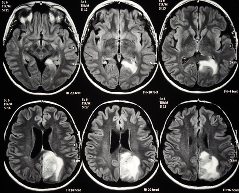
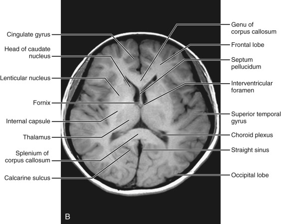
› en › e-AnatomyBrain: Atlas of human anatomy with MRI - e-Anatomy - IMAIOS Sep 13, 2021 · MRI Atlas of the Brain. This page presents a comprehensive series of labeled axial, sagittal and coronal images from a normal human brain magnetic resonance imaging exam. This MRI brain cross-sectional anatomy tool serves as a reference atlas to guide radiologists and researchers in the accurate identification of the brain structures. Brain lobes - annotated MRI | Radiology Case | Radiopaedia.org Atypical presentation of posterior reversible encephalopathy syndrome: Clinical and radiological characteristics in eclamptic patients. Aleksandra Aracki-Trenkić et al., BJBMS, 2016. A case of advanced gastrointestinal stromal tumor (GIST)presenting with paraneoplastic syndrome. H. M. Guirgis et al., J Clin Oncol, 2004. MRI head axial T2 - labeling questions - Radiopaedia The labeled structures are (excluding the correct side): cervical spinal cord posterior arch of C1 odontoid process (peg or dens) of C2 parotid gland intradural segment (V4) of dominant vertebral artery cisterna magna intradural segment (V4) of non-dominant vertebral artery cerebellar tonsil occipital condyle medulla oblongata Researchers automate brain MRI image labeling, more than ... - ScienceDaily Researchers have automated brain MRI image labeling, needed to teach machine learning image recognition models, by deriving important labels from radiology reports and accurately assigning them to ...
Brain Lesion Detection in MRI Images with Graph-cut Algorithms
A normative spatiotemporal MRI atlas of the fetal brain for automatic ... Step 2: The segmented neonatal atlases were used to generate initial labels on the spatiotemporal fetal brain MRI atlas at higher GAs (35-37 weeks) through multiatlas segmentation using probabilistic label fusion 65. Step 3: Fetal brain MRI labels were manually defined and propagated in iterations from the higher GAs to the lower GAs.

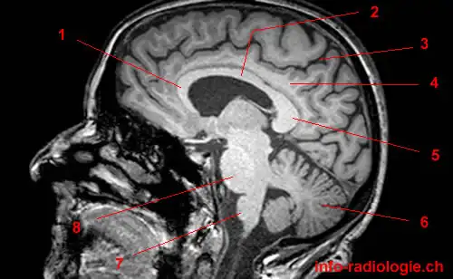
Normal anatomy of the brain on sagittal plane T1weighted ...
Brain MRI: How to read MRI brain scan | Kenhub MRI is the most sensitive imaging method when it comes to examining the structure of the brain and spinal cord. It works by exciting the tissue hydrogen protons, which in turn emit electromagnetic signals back to the MRI machine. The MRI machine detects their intensity and translates it into a gray-scale MRI image.
Normal Anatomy | Radiology Key
Atlas of BRAIN MRI - W-Radiology Brain magnetic resonance imaging (MRI) is a common medical imaging method that allows clinicians to examine the brain's anatomy (1). It uses a magnetic field and radio waves to produce detailed images of the brain and the brainstem to detect various conditions (2).
Orientation and Voxel-Order Terminology: RAS, LAS, LPI, RPI ...
academic.oup.com › brain › articleprecuneus: a review of its functional anatomy and behavioural ... Of particular interest are the neuroimaging studies seeking to define a physiological baseline state for the normal human brain function, since the precuneus shows one of the highest metabolic activity patterns of all brain regions during the conscious resting state and routinely exhibits decreases from this baseline across a variety of goal ...

Brain MRI anatomy on a sagittal T1-w... - MRI Technologist ...
› AANLIB › casesHarvard University Show labels Show list All modalities to: MR-T1 MR-T2 FDG T1/FDG T2/FDG
Design and fabrication of a realistic anthropomorphic ...
Labeled MRI Brain Scans - Neuromorphometrics The cost to label a single scan is $2449 (USD). For academic institutions, two "subscriptions" are available on-line for less than the cost of a single scan. For commercial entities, we license each neuroanatomically labeled MRI brain scans for $1500 or as an introductory special price, we'll provide every scan we currently have for ...
FDA Clears AI-Enabled Software for Streamlining Brain MRI ...
MRI Brain Atlas - University of Minnesota This web app Atlas is intended for veterinary students and radiologists seeking quick access to canine brain anatomy through a mobile device. Via a toggle button, either MRI images or approximately comparable Brain Transection images may be viewed with or without labels. Navigation & Labels.

Tips and traps in brain MRI: Applications to vascular ...
CaseStacks.com - MRI Brain Anatomy Labeled scrollable brain MRI covering anatomy with a level of detail appropriate for medical students. Show/Hide Labels. MRI Brain Anatomy. Back to Anatomy Overview. ... Labelled radiographs and CT/MRI series teaching anatomy with a level of detail appropriate for medical students and junior residents. Pelvis. Pelvic MRI anatomy

MRI Scans Show The Horrific Effect Cocaine Abuse Can Have On ...
101 Labeled Brain Images and a Consistent Human Cortical Labeling ... Another important application of labeled brain images is to train, test, and evaluate automated registration, segmentation, parcellation, and labeling algorithms. ... Automatic anatomical brain {MRI} segmentation combining label propagation and decision fusion. Neuroimage 33, 115-126 10.1016/j.neuroimage.2006.05.061 ...

Anatomy of the brain (MRI) | Mri brain, Anatomy, Mri

Mri Magnetic Resonance Imaging Brain Transverse Stock Photo ...
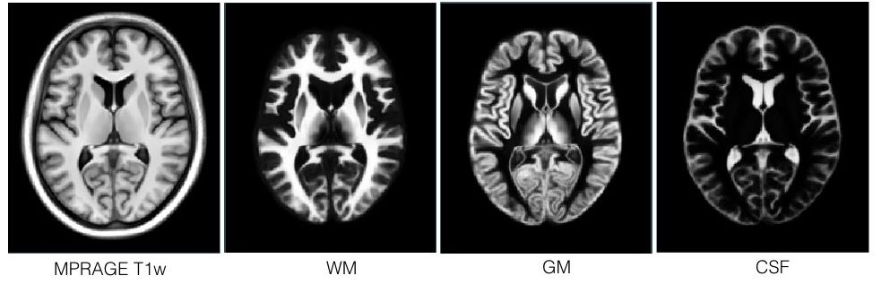
T1-weighted in vivo human whole brain MRI dataset with an ...

MRI anatomy | free MRI axial brain anatomy

Labelled Anatomy Tutorial: Axial Brain | Facebook

How to Read a MRI of Brain - Brain Anatomy MRI Explained in English

MRI anatomy | free MRI axial brain anatomy
MRI Viewer on the App Store

Radiology Reports: Reading and Understanding | AffordableMRI.com

Normal anatomy of the brain on CT and MRI with a few normal ...

Labeled MRI Brain Scans

MRI anatomy | free MRI axial brain anatomy
Evaluation of 14 nonlinear deformation algorithms applied to ...
Multi-contrast PD25 atlas – NIST

bio 151- mri human brain Diagram | Quizlet

Anatomy of the brain: T2 weighted magnetic resonance image ...

Brain Tumor Detection and Localization - Analytics Vidhya
Atlas of BRAIN MRI - W-Radiology
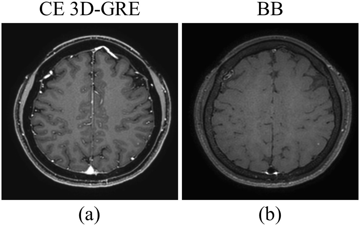
Deep-learned 3D black-blood imaging using automatic labelling ...
MRI scans prove useful for understanding depression

Brain Anatomy MRI- Neuroradiology
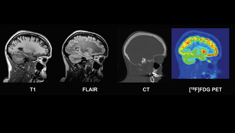
New database of healthy adult human brain PET, MRI and CT ...

Region Of Interest Based Image Classification: A Study in MRI ...

MRI anatomy | free MRI axial brain anatomy

Arterial Spin Labeling Perfusion of the Brain: Emerging ...
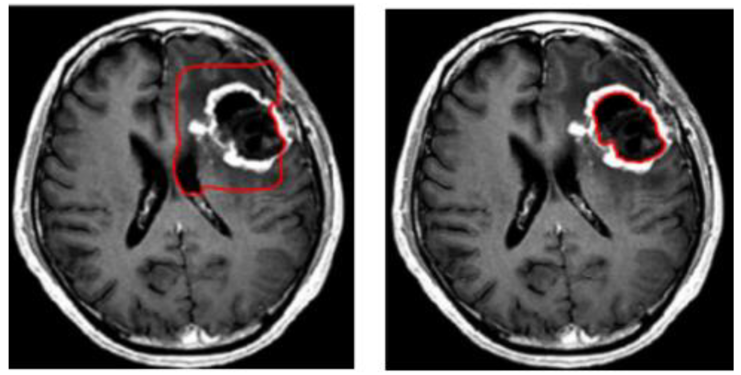
Symmetry | Free Full-Text | 3D-MRI Brain Tumor Detection ...

Spinal nerve levels, sagittal MRI - Stock Image - C030/3581 ...

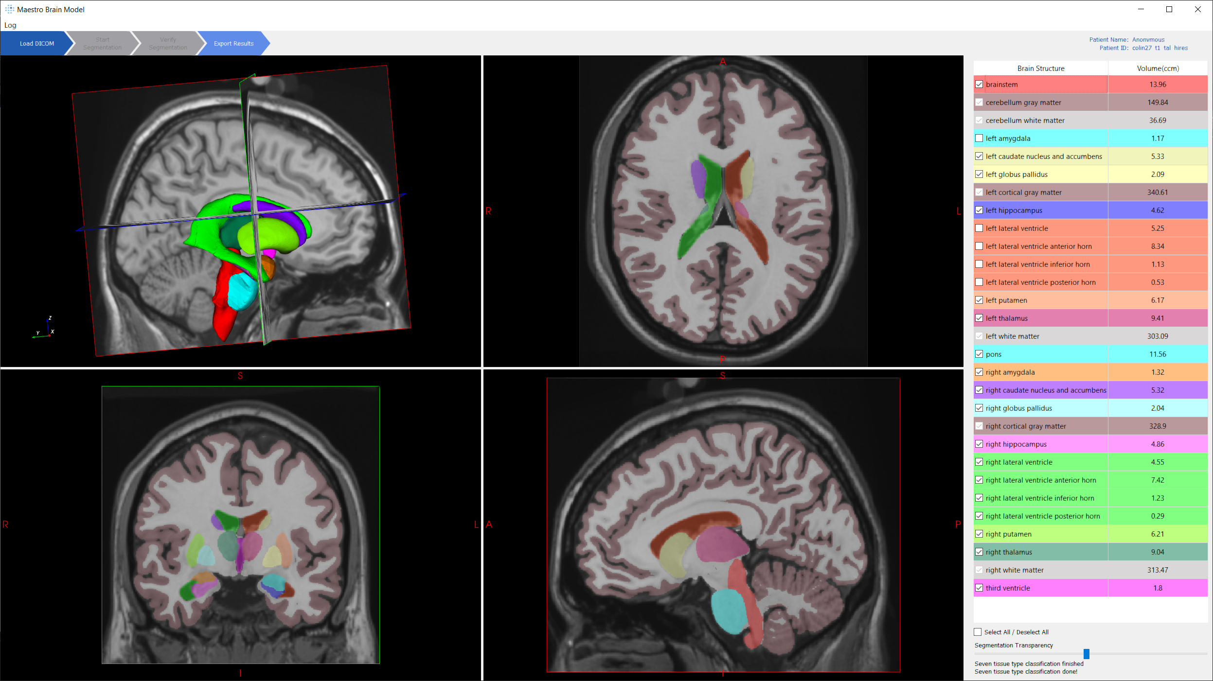
volBrain: Automated MRI Brain volumetry system

NeuroRad for iPad is a great app for medical professionals to ...
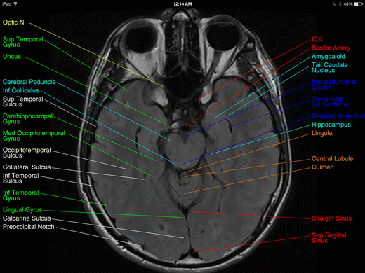
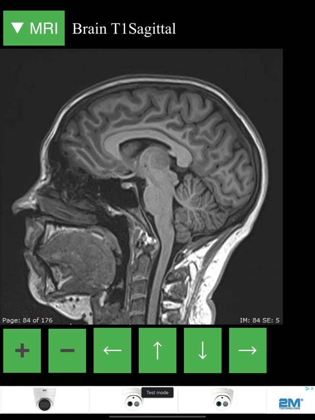
Review of “Brain MRI Atlas” App for the iPad | SpringerLink
Post a Comment for "39 brain mri with labels"